Ultrasound Engineering
Wearable Ultrasound Probe Design: From Skin Contact to Future Applications
A source-led engineering review of wearable ultrasound probe design: skin coupling, deformable arrays, beamforming, data and thermal budgets, and future applications with explicit research limitations.
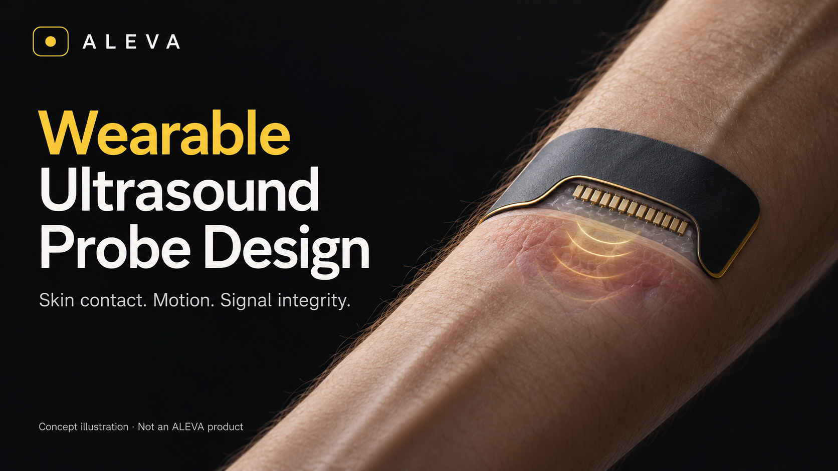
Concept illustration — not an ALEVA product or measured performance.
By ALEVA | California, USA
Engineering question: How can an ultrasound probe keep producing a trustworthy measurement when the skin, the target and the probe itself are moving?
A wearable ultrasound probe is more than a smaller handheld transducer. It must maintain an acoustic interface, tolerate body motion and operate within the power and thermal budget of a body-worn system. The important development question is not simply whether the patch bends, but whether its measurement remains interpretable after it bends.
1. Start with the measurement, not the patch
Tracking a vessel wall, imaging an organ and recognizing a hand movement are different jobs. They require different fields of view, sampling schedules and output data. Define the target depth, required observable, permitted motion, wear duration and conditions that invalidate a result before selecting an array.
A 2026 study in Nature Electronics demonstrated an integrated wireless ultrasound wristband for hand tracking and robotic or virtual-reality interaction. That is evidence for a particular sensing task, not proof that one wearable architecture can perform every diagnostic examination. [1]
2. Choose where the system should flex
Wearability does not require every layer to stretch. Wang and colleagues' 2022 bioadhesive ultrasound study combined a thin, rigid probe with a soft hydrogel-elastomer coupling interface. It demonstrated an alternative to making the entire transducer deformable. This is foundational research, not a newly launched 2026 product. [2]
A different route uses rigid sensing islands connected through compliant structures. A 2025 Nature Communications study reported flexible silicon-nanocolumn CMUT patches. Its human imaging work involved nine healthy participants and external commercial ultrasound electronics; fully integrated wireless operation was discussed as future development. A flexible transducer is therefore not, by itself, a cable-free wearable system. [3]
For an OEM design review, compare stable local element geometry with comfort and conformability. State which parts may deform, how interconnects accommodate strain, and which deformation states require recalibration or rejection.
3. Treat skin coupling as a controlled subsystem
The attachment layer is part of the acoustic path, not just packaging. The 2022 bioadhesive study explicitly engineered adhesion and resistance to dehydration into its coupling material. It also used an external acquisition system; the patch demonstration should not be described as a complete wireless product. [2]
Our proposed verification plan would examine the interface after initial placement, motion, sweating, partial lifting and the intended wear interval. Record echo stability and usable acquisition time alongside attachment condition. Distinguish the intended reusable electronics from any replaceable skin-contact component, then evaluate the actual assembly rather than an idealized water-coupled transducer.
4. Co-design the array and the beamformer
The 2023 wearable cardiac imager by Hu and colleagues combined an orthogonal transducer layout, compliant electrodes, encapsulation and imaging strategies. It illustrates that wearable imaging performance comes from the combined acoustic and system architecture, not the piezoelectric material alone. [4]
For a deformable array, our engineering recommendation is to challenge the reconstruction with the shapes expected in use. A delay model based on the undeformed element positions should not be accepted without checking the resulting image or tracking error. Validate on bent phantoms and task-relevant motion before treating a clean stationary image as sufficient evidence.
5. Budget the data path before promising wireless operation
A simple payload calculation makes the trade-off visible. Assume 32 simultaneously digitized channels, 20 million samples per second per channel and ideally packed 12-bit samples. The uncompressed sample payload is 32 × 20,000,000 × 12 = 7.68 Gbit/s before transport overhead. This is an illustrative calculation during continuous digitization, not a measured wearable device or a recommended configuration. Acquisition duty cycle and sample packing change the actual average traffic.
The design implication is to decide deliberately what leaves the patch: channel RF, processed IQ, beamformed images or task-specific features. Moving processing onto the body can reduce transmitted data but shifts computation and heat into the wearable. The 2026 technology review describes this system-level tension between processing capability, energy and form factor. [5]
6. Continuous monitoring needs quality states and exposure control
Longer operation is not automatically better operation. FDA notes that ultrasound introduces energy into tissue and can cause thermal and mechanical effects; it recommends minimizing exposure while preserving diagnostic quality under the ALARA principle. A small enclosure does not remove the need to assess acoustic exposure. [6]
We propose evaluating electronics temperature separately from acoustic output, and defining observable states such as valid acquisition, uncertain contact, target lost and temperature-limited operation. A failed measurement should be identifiable in the record, rather than silently converted into a plausible physiological value. These are proposed engineering requirements, not results from an ALEVA prototype.
7. Where future applications are becoming credible
Cardiovascular monitoring: wearable cardiac imaging has been demonstrated in research. Translation to a particular care pathway still requires evidence for its intended users, population and operating conditions. [4]
Supervised prenatal monitoring: a May 2026 Nature Biotechnology study reported autonomous vessel tracking and comparison with clinical ultrasound in 62 pregnancies. The authors identified miniaturized wireless integration as further development. This should not be presented as proof of unrestricted, unsupervised home use. [7]
Human-machine interfaces and future rehabilitation research: the 2026 wrist-imaging study demonstrated hand tracking for robotic and virtual-reality control. Using similar sensing principles to assess rehabilitation progress is a possible research direction, not an established therapeutic benefit from that demonstration. [1]
The OEM/ODM decision
Start with one well-defined task and specify the whole measurement chain: transducer, coupling layer, attachment, electronics, reconstruction and quality flags. Agree on acceptance tests for placement variation, motion, sustained operation and invalid-data handling before freezing the design. Connect those tests to a controlled manufacturing and calibration record.
The future opportunity is not simply an ultrasound patch that can be worn. It is a task-specific system that can show when its measurement is reliable—and when it is not. This article is a public-source engineering analysis; it reports no ALEVA wearable product, measured performance, regulatory authorization or clinical outcome.
Related ALEVA guide: Custom Ultrasound Probe Design.
References
Lu, G. et al. Hand tracking using wearable wrist imaging. Nature Electronics 9, 426–440. Published March 25, 2026. Peer-reviewed research; publisher abstract and MIT's author-institution report checked, not a complete subscription-paper download.
Wang, C. et al. Bioadhesive ultrasound for long-term continuous imaging of diverse organs. Science 377, 517–523. July 29, 2022; online July 28. Peer-reviewed foundational research.
Kang, D.-H. et al. Silicon nanocolumn-based disposable and flexible ultrasound patches. Nature Communications 16, 6609. July 18, 2025. Peer-reviewed open-access research.
Hu, H. et al. A wearable cardiac ultrasound imager. Nature 613, 667–675. January 25, 2023. Peer-reviewed foundational research; the DOI's 2022 identifier is not its publication date.
Yan, M. et al. Wearable ultrasound: a review of core technologies and clinical applications in cardiovascular monitoring. Microsystems & Nanoengineering 12, 246. June 23, 2026. Review article, not a new device trial.
US Food and Drug Administration. Ultrasound Imaging. Official information page, accessed October 21, 2026.
Park, G. et al. Fetal monitoring for high-risk pregnancies using a wearable ultrasound patch. Nature Biotechnology. May 26, 2026. Peer-reviewed research.